Key Knowledge:
|
The plasma membrane separates the internal components of a cell from the external environment via two key properties:
- It is semi-permeable – some material cannot cross the membrane without assistance
- It is selective – the cell can regulate the passage of these materials according to need
Membrane Structure
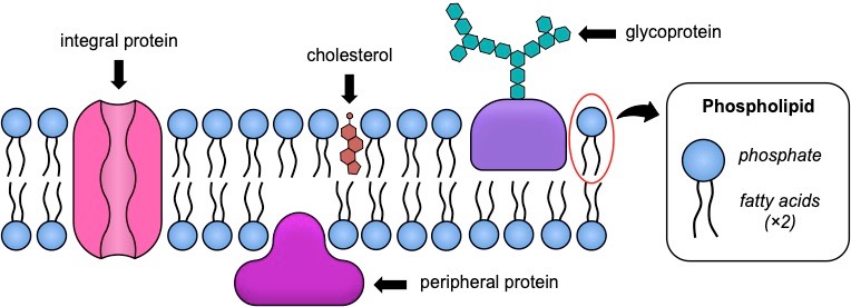
Plasma membranes consist of three principal components – a phospholipid bilayer, proteins and cholesterol (in animal cells only)
The organisation of these components is represented by the fluid-mosaic model, which reflects the fact that membranes are:
- Fluid – the phospholipid bilayer is viscous (due to weak hydrophobic associations), meaning membrane components can move position
- Mosaic – the phospholipid bilayer is embedded with proteins (and potentially cholesterol), resulting in a mosaic of components

1. Phospholipid Bilayer

Structure of Phospholipids
|
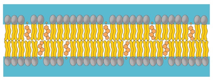
Arrangement in Membranes
|
Properties of the Phospholipid Bilayer
|
2. Membrane Proteins
Phospholipid bilayers are embedded with proteins, which may be either permanently or temporarily attached to the membrane
- Integral proteins are permanently attached to the membrane and are typically transmembrane (they span across the bilayer)
- Peripheral proteins are temporarily attached by non-covalent interactions and associate with one surface of the membrane
Membrane proteins can serve a variety of key functions, including:
- Transport: Membrane proteins can facilitate the passive or active movement of molecules that cannot freely cross the bilayer
- Metabolic Activity: Enzymes can be bound to the membrane to localise activity and receptors can detect signalling molecules
- Connections: Membrane proteins can join cells together or act as an attachment point for extracellular or intracellular components

3. Cholesterol
Cholesterol is a component of animal cell membranes, where it functions to maintain integrity and mechanical stability
- It is absent in plant cells, as these plasma membranes are surrounded and supported by a rigid cell wall made of cellulose
- Cholesterol is an amphipathic molecule (like phospholipids), meaning it has both hydrophilic and hydrophobic regions
Cholesterol interacts with the fatty acid tails of phospholipids to moderate the properties of the membrane:
- Cholesterol functions to immobilise the outer surface of the membrane, reducing fluidity
- It makes the membrane less permeable to very small water-soluble molecules that would otherwise freely cross
- It functions to separate phospholipid tails and so prevent crystallisation of the membrane
- It helps secure peripheral proteins by forming high density lipid rafts capable of anchoring the protein

Membrane Transport
Movement of materials across a membrane will depend on both the size and solubility of the material in question
- Small, lipophilic molecules can freely pass across the phospholipid bilayer (e.g. oxygen, carbon dioxide, water, steroids)
- Larger molecules (e.g. glucose) or polar / charged molecules (e.g. ions) will require membrane proteins in order to cross
Passive transport involves the movement of material along a concentration gradient (high concentration ⇒ low concentration)
- Because materials are moving down a concentration gradient, it does not require the expenditure of energy (ATP hydrolysis)
- There are three main types of passive transport mechanisms: simple diffusion, facilitated diffusion or osmosis
Active transport involves the movement of materials against a concentration gradient (low concentration ⇒ high concentration)
- Because materials are moving against the gradient, it requires the expenditure of energy (e.g. ATP hydrolysis)
Bulk transport (cytosis) involves materials entering or leaving the cell via vesicles (the membrane breaks and reforms)
- In this form of transport, materials do not cross the membrane directly and can move along or against the gradient
- This is an active process (requires ATP) but it is not active transport (as materials are circumventing the membrane)
1. Simple Diffusion
Simple diffusion involves the net movement of molecules from a region of high concentration to a region of low concentration
- This directional movement along a gradient is passive and will continue until molecules become evenly dispersed (equilibrium)
- Small and non-polar (lipophilic) molecules will be able to freely diffuse across cell membranes (e.g. O2, CO2, glycerol)
The rate of diffusion can be influenced by temperature (affects kinetic energy), molecular size and the size of the concentration gradient
2. Facilitated Diffusion
Facilitated diffusion is the passive movement of molecules across the cell membrane via the aid of a membrane protein
- It is utilised by molecules that are unable to freely cross the phospholipid bilayer (e.g. large, polar molecules and ions)
This process is mediated by two distinct types of transport proteins – channel proteins and carrier proteins
- Channel proteins have a hydrophilic pore and may be gated to regulate the passage of ions in response to certain stimuli
- Carrier proteins undergo a conformational change to translocate solutes and have a comparably slower rate of transport

3. Osmosis
Osmosis is the net movement of water molecules across a semi-permeable membrane from a region of low solute concentration to a region of high solute concentration (until equilibrium is reached)
- Water is considered the universal solvent – it will associate with, and dissolve, polar or charged molecules (solutes)
- Because solutes cannot cross a cell membrane unaided, water will move to equalise the two solutions
- At a higher solute concentration there are less free water molecules in solution as water is associated with the solute
- Osmosis is essentially the diffusion of free water molecules and hence occurs from regions of low solute concentration

Solutions may be loosely categorised as hypertonic, hypotonic or isotonic according to their osmotic effect
- Solutions with a relatively higher solute concentration are categorised as hypertonic (and will gain water)
- Solutions with a relatively lower solute concentration are categorised as hypotonic (and will lose water)
- Solutions that have the same solute concentration are categorised as isotonic (there will be no net water flow)
4. Active Transport
Active transport is the active movement of molecules across the cell membrane via the aid of a protein pump
- Active transport is essentially the reverse of facilitated diffusion (it moves materials against a concentration gradient)
- The pump binds a solute and then undergoes a conformational change to translocate solutes across the membrane
- The conformational change is mediated by the hydrolysis of ATP (to ADP + Pi) and is hence energy dependent

5. Bulk (Vesicular) Transport
The fluidity of membranes allows materials to be taken in or released by cells without crossing the phospholipid bilayer
- The membrane is principally held together by weak hydrophobic associations between the fatty acid tails of the phospholipids
- These weak interactions can be spontaneously broken and reformed in a process that is energy dependent (via ATP hydrolysis)
- Membrane segments can be excised to form internal vesicles, while new segments are added when membrane and vesicle fuse
Endocytosis
Endocytosis involves large substances (or bulk amounts of smaller substances) entering the cell without crossing the membrane
- The membrane forms a flask-like depression (i.e. invagination) which envelopes the extracellular material to be internalised
- The invagination is then sealed off to form an intracellular vesicle containing the material

There are two main types of endocytosis:
- Phagocytosis – The process by which solid substances are ingested (usually to be transported to the lysosome)
- Pinocytosis – The process by which liquids / dissolved substances are ingested (allows faster entry than via protein channels)
Exocytosis
Exocytosis involves substances exiting the cell without crossing the membrane (these substances must be packaged in vesicles)
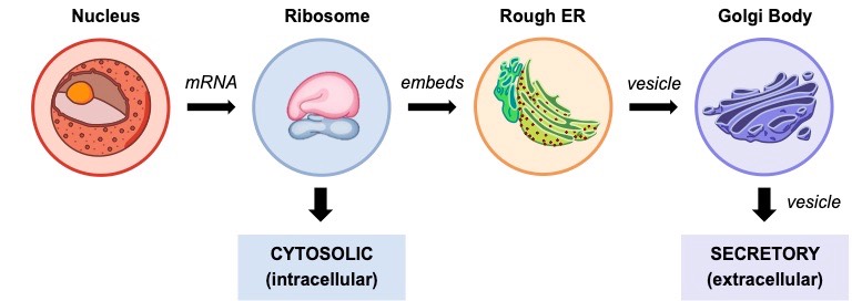
- Polypeptides synthesised within the rough ER will be packaged into a vesicle and transported to the Golgi apparatus
- The Golgi complex will potentially modify the protein (e.g. glycosylation) and then export it out of the cell via a vesicle (exocytosis)
- Proteins can either be secreted by the Golgi immediately (constitutive secretion) or stored for a delayed release (regulatory secretion)
- Proteins synthesised via this pathway can also be shuttled to other organelles besides the Golgi (e.g. mitochondria, lysosome, etc.)
